

A 6-year-old male presented to the pediatric emergency department (PED) for scalp lesions. He was seen by his pediatrician 2 weeks prior and prescribed antibiotics and a delousing shampoo for suspected cellulitis versus lice infestation. Symptoms did not improve despite completion of treatment. An outpatient ultrasound was performed showing “multiple scalp echogenic nodular lesions measuring from 0.5 cm to 1.2 cm in the long axis diameter.” The following differential diagnosis was entertained: lymphadenitis, benign avascular mass, epidermal inclusion cyst, or pilomatricoma, and the patient was started on clindamycin. Due to concern for an oncologic process, a surgery consultation was placed to arrange for a biopsy. Four days after the ultrasound and before the biopsy could be performed, the patient and his mother presented to the PED due to worsening symptoms. Multiple new lesions developed across the patient’s scalp which bled when pressure was applied. The patient denied fever and reported intermittent pruritus and pain over the lesion sites. The mother reported a history of travel to Ecuador one month prior to symptom onset.


Vitals: BP 98/61; Pulse 73; Temp 36.3°C (97.3°F) temporal; Resp 18; SpO2 99%, RA
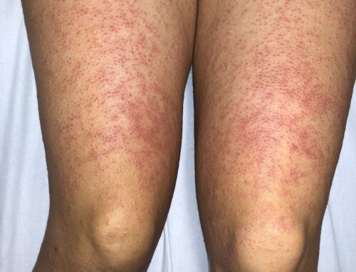
Skin: Large, 3 x 3cm indurated, erythematous lesion located over the patient’s right temporal scalp (Image 1). Five additional lesions noted across the entirety of the scalp. No lesions identified below the neck. Lesions are mildly tender to palpation; no fluid able to be expressed. A small centrally located pore is noted on each lesion with appearance of pulsatile fluid level. No associated lymphadenopathy. A point-of-care ultrasound (POCUS) using a high-frequency, linear transducer was performed during the PED visit (Image 2).
Non-contributory
In short axis, there is an echogenic lesion with surrounding fluid (halo sign) suggesting a foreign body that also exhibits posterior acoustic shadowing. With the transducer held still, independent movement is visualized within the center of the lesion (Image 3).
Cutaneous furuncular myiasis due to Dermatobia hominis (botfly infestation).
Take-Home Points
- Native to Central and South America, botfly infestation is facilitated through an infected female mosquito which deposits its eggs on the skin of a mammal on which it feeds.
- Cutaneous furuncular myiasis is important to consider for unexplained head, neck, and extremity lesions when there is suspected travel to endemic areas and is unlikely to be recognized in the continental United States due to low prevalence.
- Consider pertinent physical exam findings and utility of POCUS in confirming the diagnosis.
- Harris AT, Bhatti I, Bajaj Y, Smelt GJ. An unusual cause of pre-auricular swelling. J Laryngol Otol. 2010 Mar;124(3):339-40. doi: 10.1017/S002221510999082X. Epub 2009 Aug 11. PMID: 19664319.
- Minakova E, Doniger SJ. Botfly larva masquerading as periorbital cellulitis: identification by point-of-care ultrasonography. Pediatr Emerg Care. 2014 Jun;30(6):437-9. doi: 10.1097/PEC.0000000000000156. PMID: 24892687.

Eric Boccio, MD
Instructor
UMass Chan Medical School – Baystate
Latest posts by Eric Boccio, MD (see all)
- SAEM Clinical Images Series: Unusual Scalp Lesions - October 2, 2023
