Benign Skin Lesions
Introduction
A skin lesion is a part of the skin that has an abnormal growth or appearance compared to the skin around it.
Common benign skin lesions have typical clinical characteristics and appearances.
- Certain lesions, such as nevi, milia, dermatofibroma, skin tags, pyogenic granuloma, keratosis pilaris, syringoma are more common in children and younger adults.
- Some lesions such as seborrheic keratosis, sebaceous hyperplasia, solar lentigo, cherry angioma, large cell acanthoma, venous lake, ganglion, epidermal inclusion cysts, and lipoma are more likely to present in middle-aged to older adults.
Benign lesions that are symptomatic or cosmetically bothersome can often be managed with simple procedures such as cryotherapy, electrosurgery or excision.
ABCDE Criteria
All skin lesions should be assessed for signs that may suggest malignancy such as newly changing appearance or any positive ABCDE criteria.
- A - Asymmetry
- B - Border irregularities
- C - Colour variation (mottled, shades of brown, black, grey and white)
- D - Diameter >6 mm (size of a pencil eraser)
- E - Evolving size, shape, surface (raised, bleeding, crusting), shades of colour, or symptoms (itchiness, tenderness)
Milia
Milia are very small (≤3 mm in size), raised, pearly-white or yellowish bumps on the skin. It is seen frequently in newborn infants, typically on face, scalp, upper trunk and upper extremities, and resolve spontaneously within few weeks to months. However, when arises in adults, it may be persistent.
Typically, milia do not usually need any treatment. However, milia can be treated for cosmetic reason by incision of the overlying epidermis and expression of the content. Smaller lesions may respond to topical retinoids applied daily for several weeks.
NOTE: Milia should be distinguished from comedones, xanthelasma and syringomas.
Skin Tag
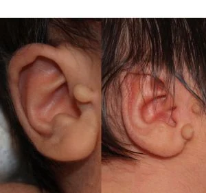
Acrochordons, commonly known as skin tags, are an outgrowth of normal skins (soft and fleshy).
- They are frequently seen in obese patients and in patients with diabetes mellitus. Observational studies suggest that acrochordons may be a skin sign of insulin resistance and metabolic syndrome.
- Acrochordons usually occur in sites of friction, particularly the axilla, neck, inframammary, and inguinal regions. They become symptomatic when traumatized (e.g., caught on jewellery, rubbed by clothing).
- Treatment is indicated if lesions are irritating or the patient desires removal for cosmetic reasons.
Solar Lentigines
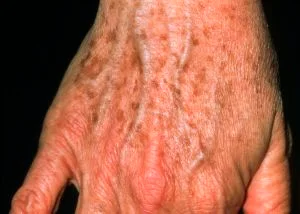
Solar lentigines typically appears as new lesion (harmless patch of darkened skin) in patient aged >50 years (commonly called "age spots" or "liver spots").
- It results from exposure to ultraviolet (UV) radiation, which causes proliferation of melanocytes and accumulation of melanin within the skin cells (keratinocytes).
- Preventive measurements include minimizing sun exposure and use of sunscreens, but this needs to start early in life.
External Links
- DynaMed - Common Benign Skin Lesions
- UpToDate - Overview of Benign Skin Lesions of the Skin
- AFP - Diagnosing Common Benign Skin Tumors, 2015
- Milia en plaque: a new site, 2000
- Milia: a review and classification, 2008
- Sun-induced freckling: ephelides and solar lentigines, 2014
- Acrochordons as a cutaneous sign of metabolic syndrome: a case-control study, 2014


Comments
Post a Comment