

An 86-year-old man with a past medical history of coronary artery disease, hypertension, hyperlipidemia, chronic kidney disease, COPD, choledocholithiasis requiring ERCP and sphincterotomy 2 years ago presented with five days of feeling unwell. History was limited due to cognitive impairment. His daughter had reported to staff he had been feeling unwell for five days, intermittently having nausea and generalized abdominal pain, subjective fevers, chest pain, and shortness of breath. His daughter also reported a history of intermittent lower abdominal cramping which was chronic for him. He denied changes to urination or bowel movements.


Vitals: BP 106/67, Temp 36.2°C, Pulse 115, Resp 20, SpO2 95%
General: Nontoxic appearing, no distress
Heart: Regular, no murmurs
Lungs: Clear bilaterally, normal work of breathing
Abdomen: Diffusely tender, greatest in left upper quadrant
CBC with differential: WBC 14.1, Neutrophil 12% (high)
Comprehensive metabolic panel (CMP): Total bilirubin 2.7 (high), AlkP 328 (high), AST/ALT normal
Lipase: Normal
Troponin x2: Negative
Chest x-ray: No acute abnormality
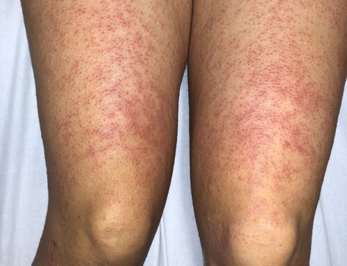
This patient has sonographic evidence of perforated gangrenous cholecystitis which was confirmed on subsequent CT scan. Gallbladder perforation is a complication of cholecystitis and has a reported incidence of 5-10%. It has been reported as early as two days after the onset of symptoms to as late as several weeks afterward. The most common site of perforation is the fundus due to relatively poor blood supply. In this case, the culprit perforation was in the proximal body adjacent to the stone which is suspected to have eroded through the wall.
Figure 1 depicts a minimally thickened gallbladder wall measured at 3.5 mm with a large shadowing stone-in-neck and associated perihepatic fluid collection (arrow) with a subtle intraluminal membrane and wall irregularity consistent with gangrenous cholecystitis. Figure 2 doppler images show no flow within the fluid collection and a suspiciously thin gallbladder wall (arrow). Figure 3 again highlights an irregular wall with small “hole sign” (arrow) signifying perforation of the gallbladder into the adjacent fluid collection. This patient was admitted to the hospital’s general surgical service and treated with IV broad-spectrum antibiotics and a percutaneous cholecystostomy tube placed by interventional radiology.
Take-Home Points
- Look out for “hole signs” with adjacent fluid collection on your gallbladder ultrasounds which would suggest perforation.
- Intraluminal membranes or wall irregularities suggest gangrenous cholecystitis.
- Initial treatment includes broad-spectrum antibiotics and cholecystostomy tube decompression.
- Indiran, V., Prabakaran, N. & Kannan, K. “Hole sign” of the gallbladder. Indian J Gastroenterol 36, 66–67 (2017). https://doi.org/10.1007/s12664-016-0723-3
- Jeffrey RB, Laing FC, Wong W, Callen PW. Gangrenous cholecystitis: diagnosis by ultrasound. Radiology. 1983 Jul;148(1):219-21. doi: 10.1148/radiology.148.1.6856839. PMID: 6856839.
- Sood, B.P., Kalra, N., Gupta, S., Sidhu, R., Gulati, M., Khandelwal, N. and Suri, S. (2002), Role of sonography in the diagnosis of gallbladder perforation. J. Clin. Ultrasound, 30: 270-274.

Jonathan Woo, MD
Stanford Health Care
Latest posts by Jonathan Woo, MD (see all)
- SAEM Clinical Images Series: An Ultrasonographic Rabbit Hole - February 16, 2024